


Applications in Industry and Science
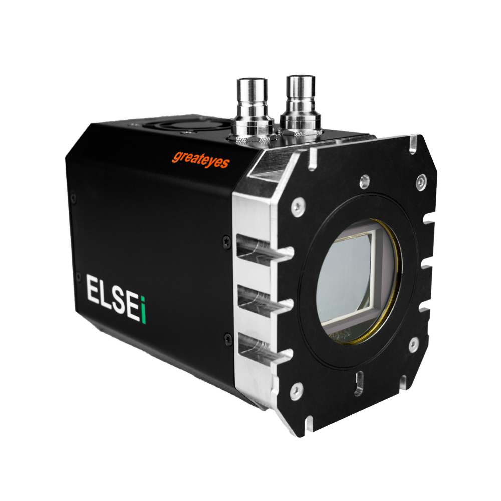
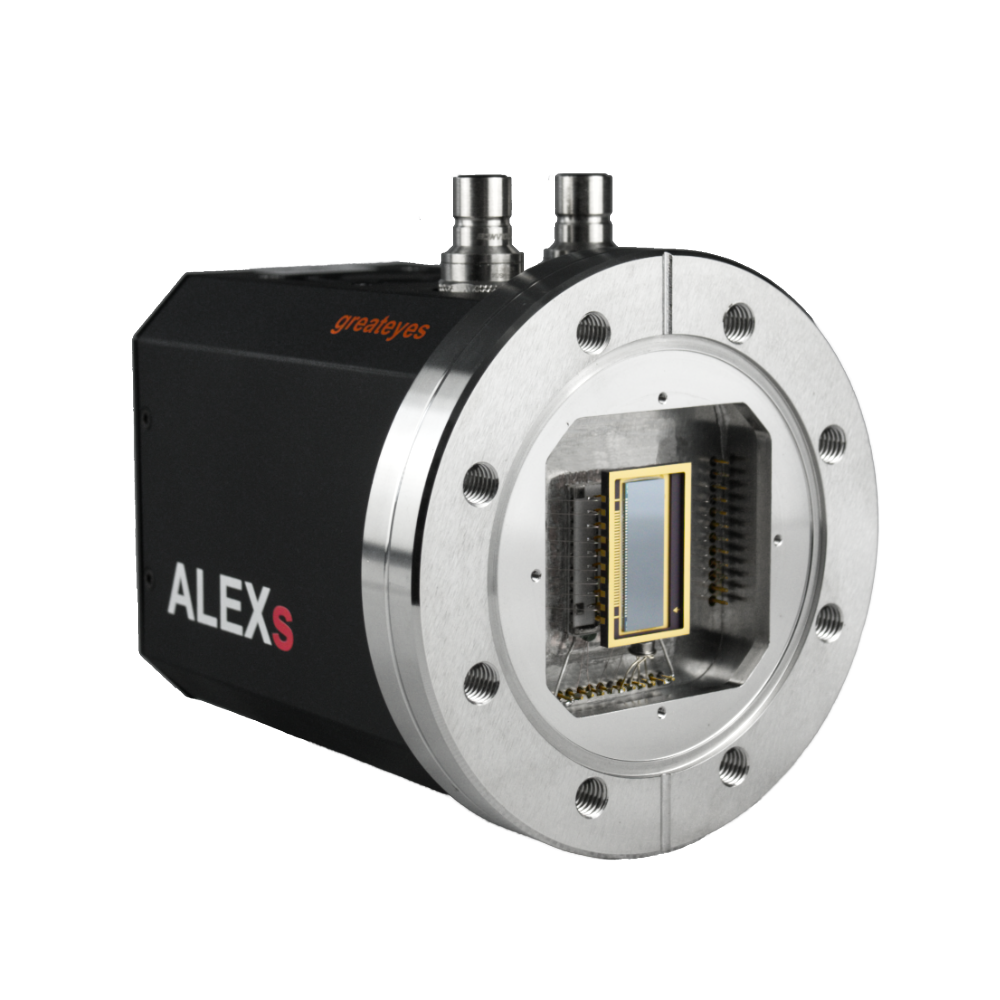
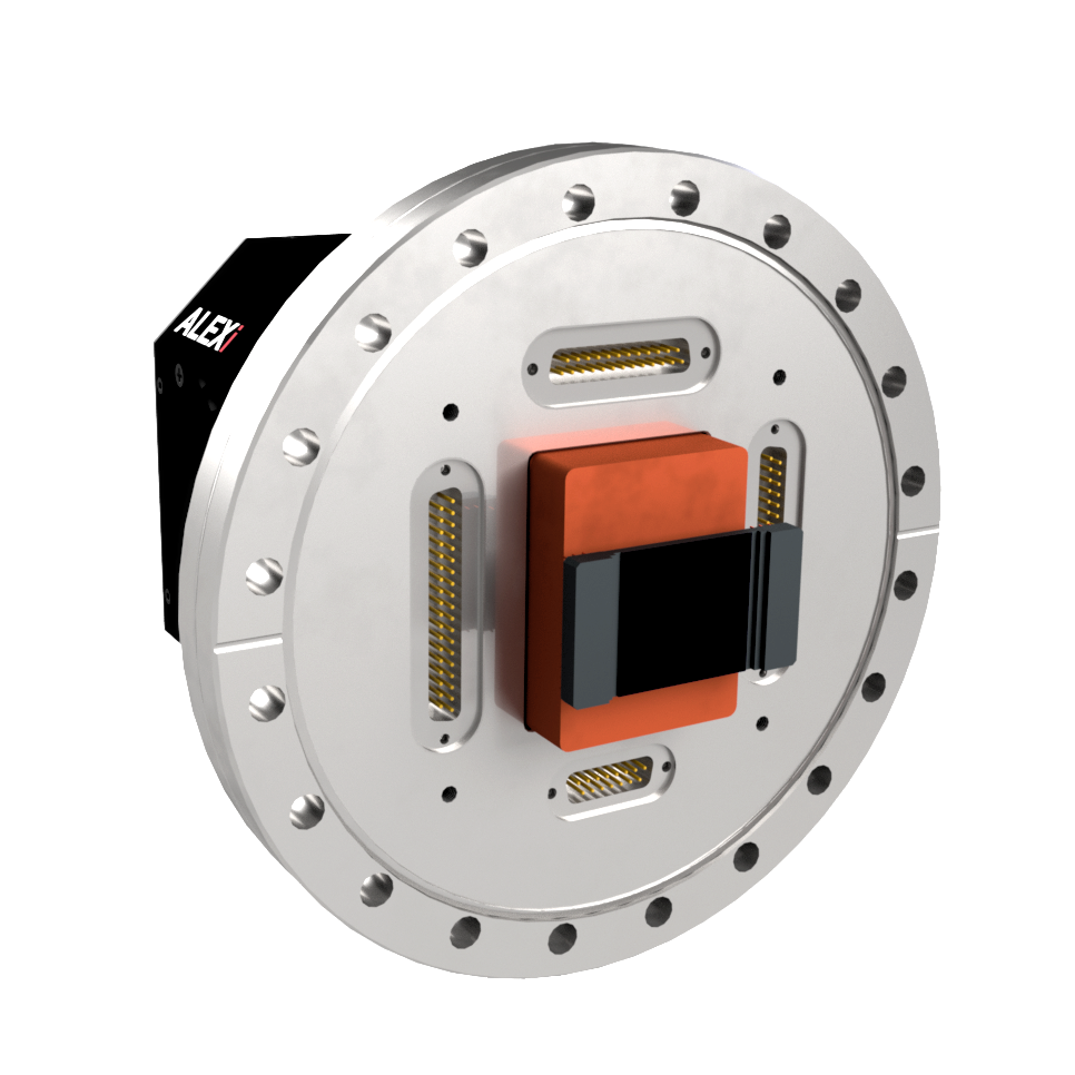
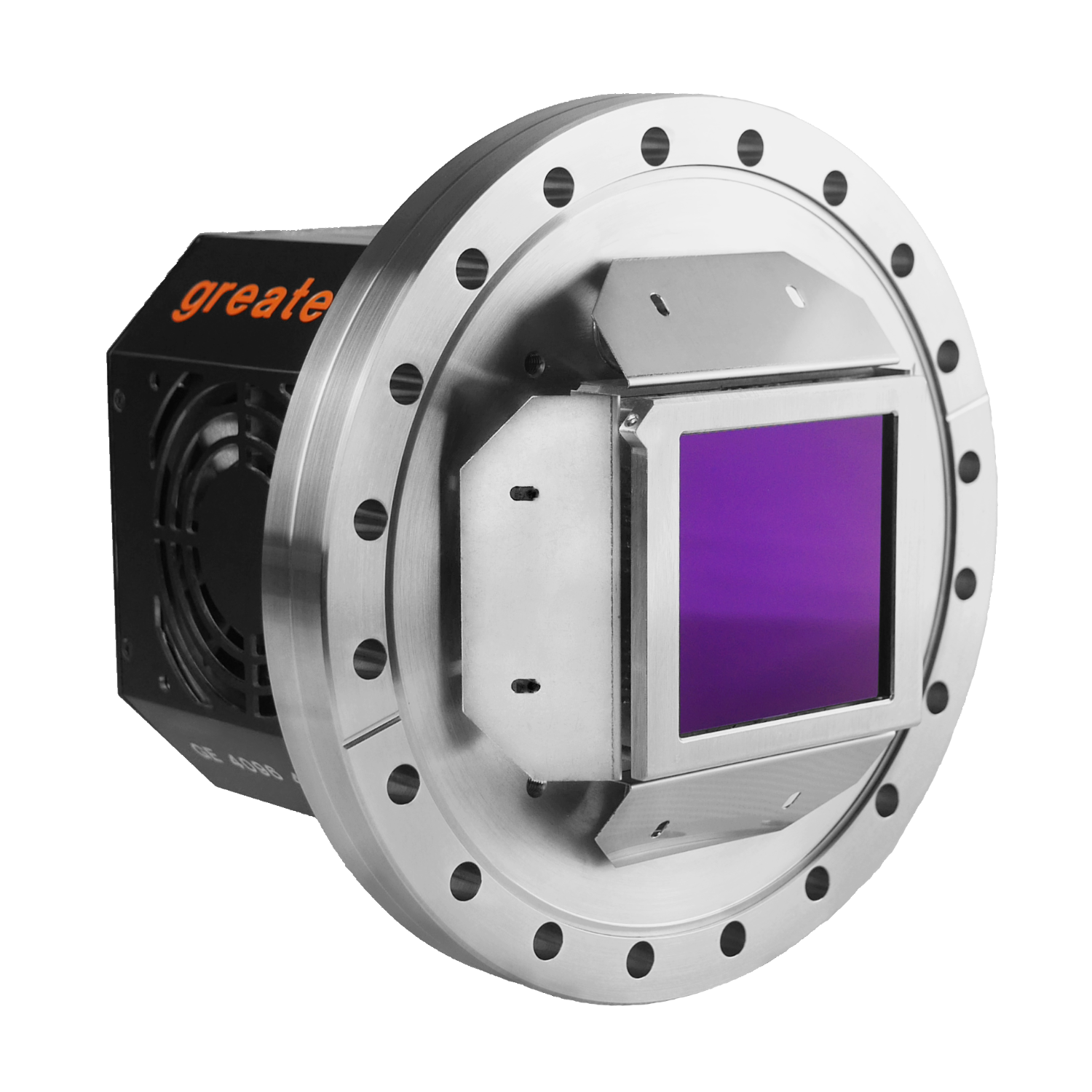
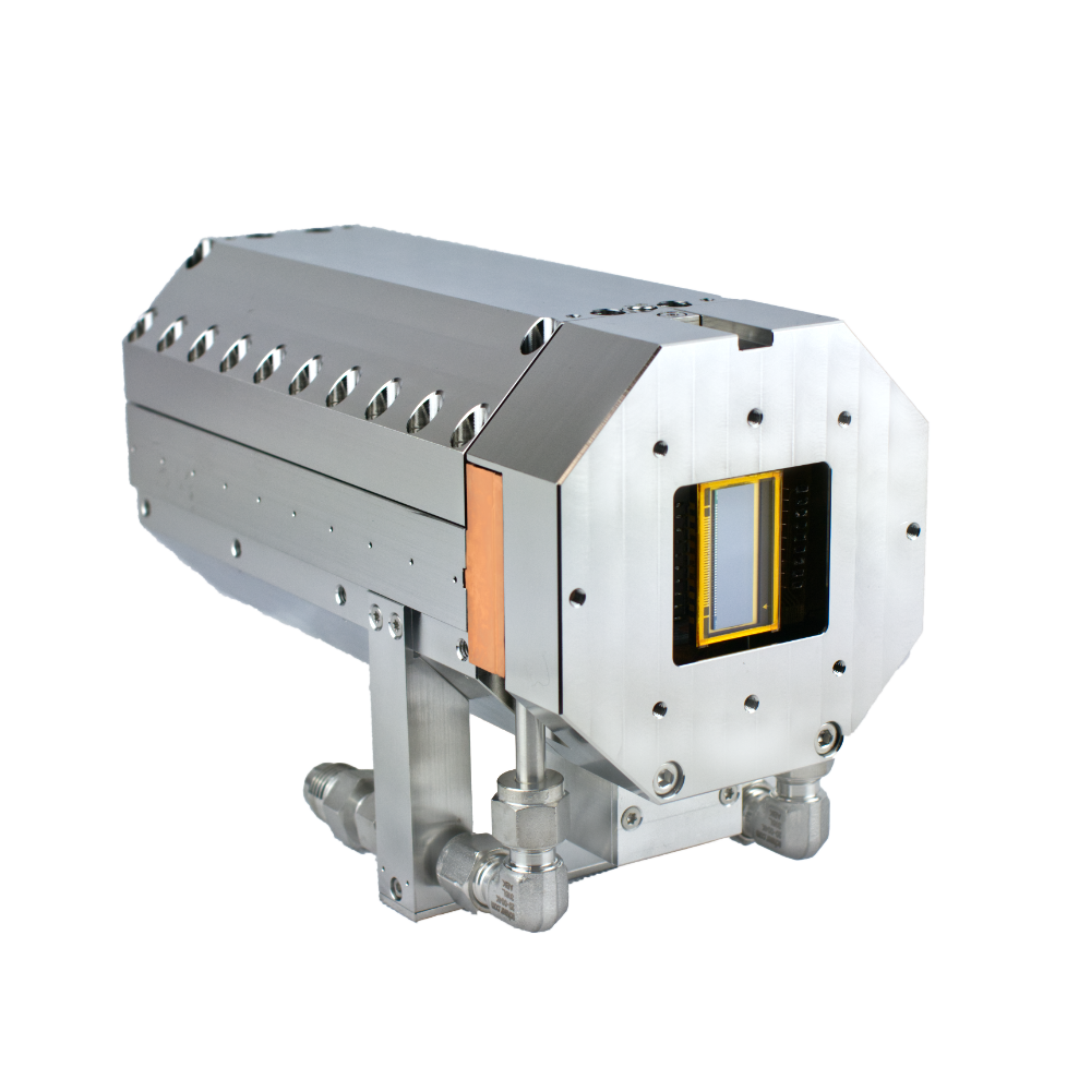
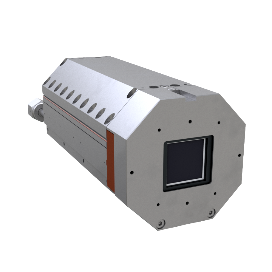
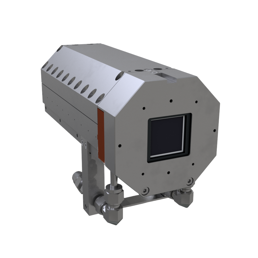
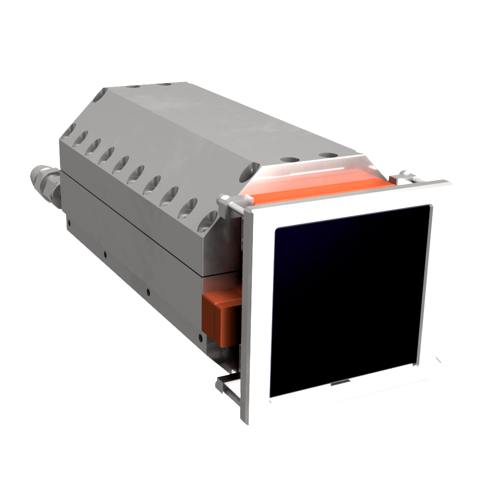
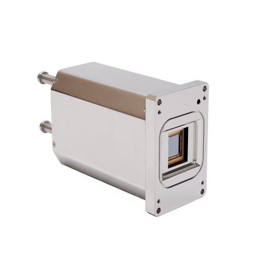
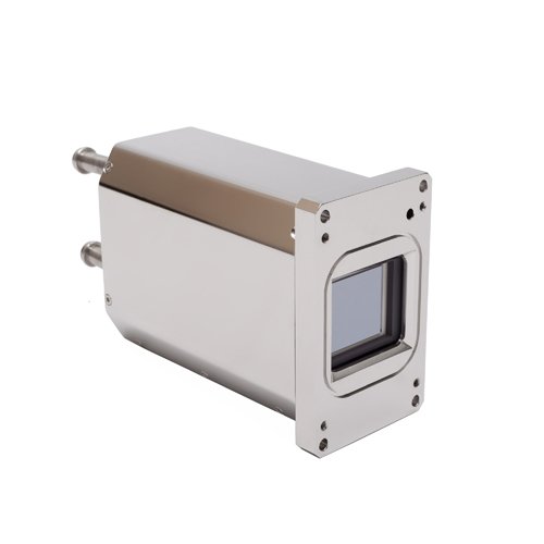
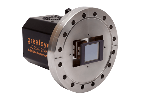
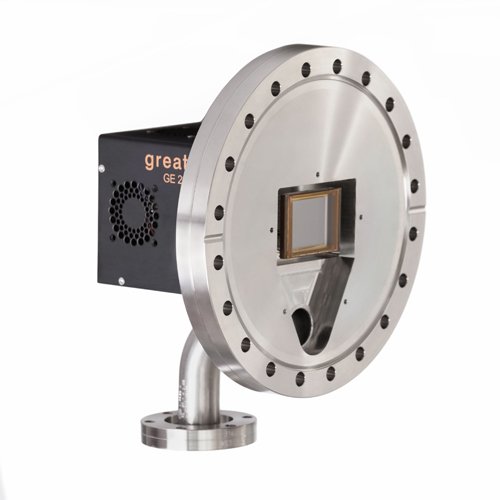
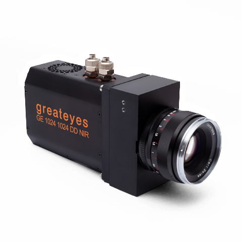
Scientific high-sensitive cameras cameras from greateyes are qualified for imaging and spectroscopy from NIR, VIS, UV-range to EUV- and Xray-range. They combine highly sensitive sensors with ultra low noise electronics for optimal detection of weak signals. The following examples show a selection of applications with greateyes cameras involved.Get in touch with our experts to find the most qualified greateyes camera for your application.
| Spectroscopy | |
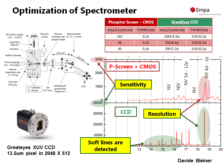 |
Detectors for XUV Spectroscopy: Phosphor-Screen camera versus greateyes XUV camera greateyes XUV cameras deliver much higher sensitivity and spectral resolution in comparison to Phosphor-Screen based cameras systems. Thanks to Dr. Davide Bleiner, EMPA. |
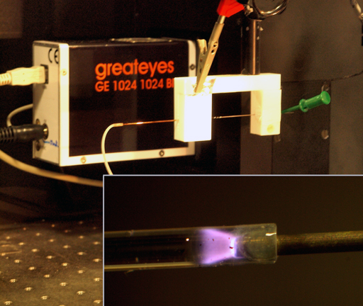 |
Plasma Spectroscopy and Emission Spectroscopy At ISAS Dortmund a miniaturized plasma with a liquid electrode (Liquid Electrode Dielectric Barrier Discharge, LE-DBD) has been developed which can be operated in continous flow operation requiring only small sample volumes. |
 |
EUV transmission spectroscopy The method of EUV transmission spectroscopy provides a fast thickness characterization for thin foils and is based on a laser induced plasma source which emits light in the EUV. |
 |
X-Ray Imaging and X-Ray Spectroscopy At the Berlin Laboratory for innovative X-ray technologies (BLIX) - Technical University Berlin a modular Laser-Plasma Source for Spectroscopy emitting Soft X-Ray radiation is being developed. |
| Imaging | |
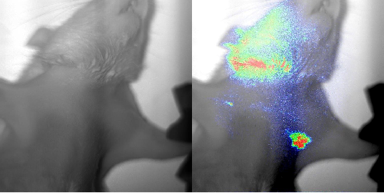 |
Fluorescence in vivo imaging Fluorescence in vivo imaging works on the basis of fluorochromes that are excited by an external light source, and which emit light of a different wavelength in response. The emitted fluorescence can be detected by using a camera which is sensitive in the spectral range of near-infrared. |
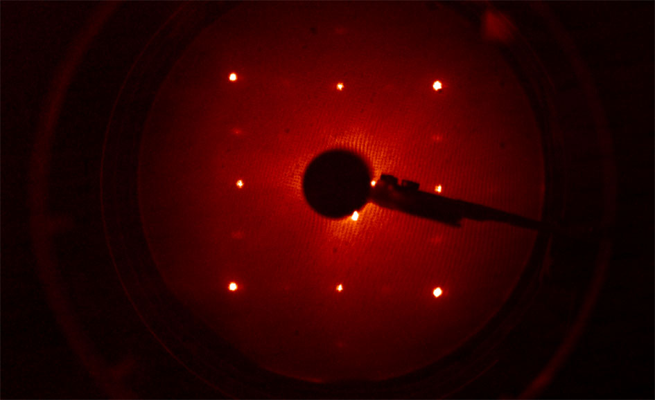 |
LEED imaging Low Energy Electron Diffraction is a technique used to determine the atom formation on surface structures and thin films of crystalline materials. |
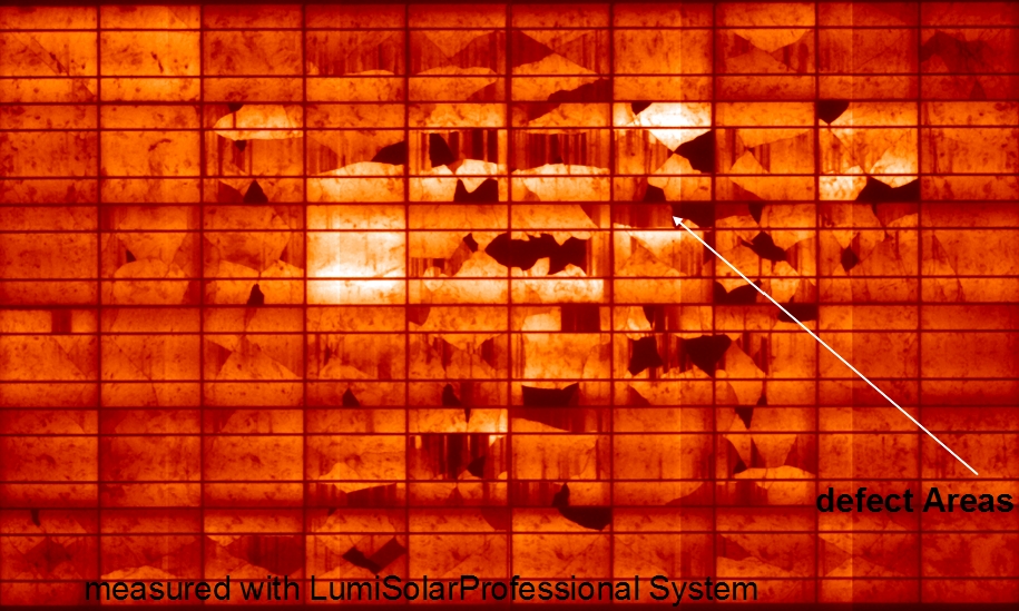 |
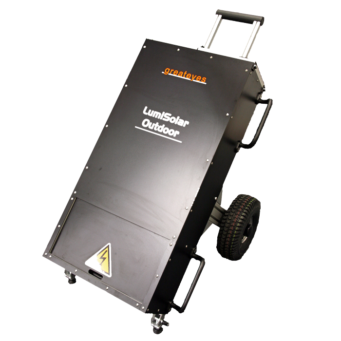
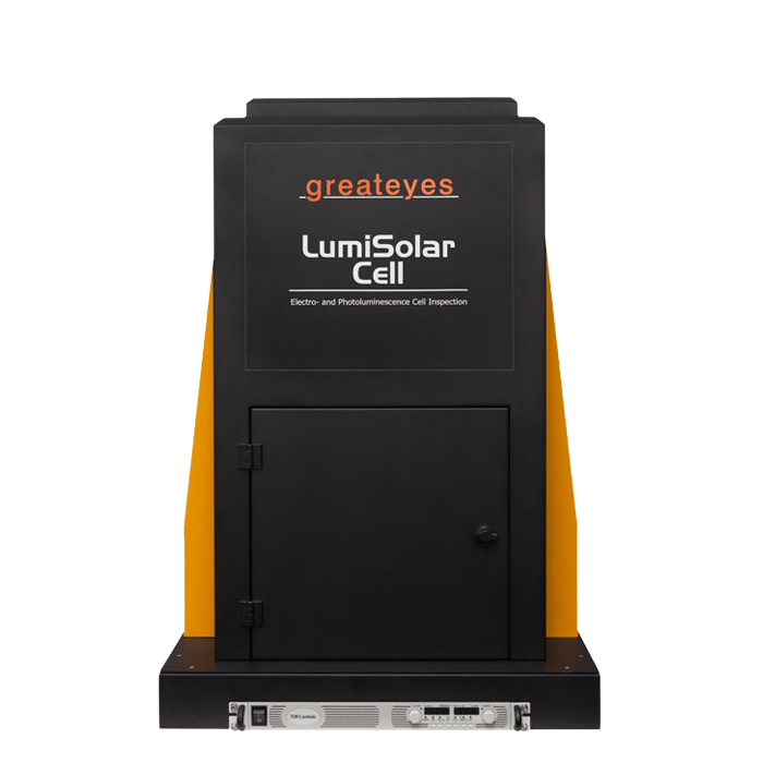
Electroluminescence An external current is applied to the solar cell or module using its electrical contacts. The invisible electroluminscence radiation emitted by the solar cell or module is detected by a highly sensitive camera. |
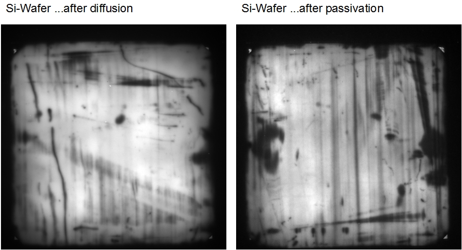 |
Photoluminescence The solar cell or wafer is excited by an intensive light source. No electrical connections to the solar cell are necessary. The invisible photoluminescence radiation emitted by the wafer or solar cell is detected by a highly sensitive camera. |
| Additional examples | |
| Chemiluminescence | Fluorescence Spectroscopy |
| Electrochemiluminescence | Phosphorescence Spectroscopy |
| Bioluminescence | Laser induced Breakdown Spectroscopy |
| Biochip Reading | Raman Spectroscopy |
| Gel Scanner | Atom Absorption Spectroscopy |
| Transient Absorption Spectroscopy | |
Selected references:
Julius Reinhard, Sophia Kaleta, Johann Jakob Abel, Felix Wiesner, Martin Wünsche, Eric Seemann, Martin Westermann, Thomas Weber, Jan Nathanael, Alexander Iliou, Henryk Fiedorowicz, Falk Hillmann, Christian Eggeling, Gerhard G Paulus, Silvio Fuchs, Laboratory-Based Correlative Soft X-ray and Fluorescence Microscopy in an Integrated Setup, Microscopy and Microanalysis, Volume 29, Issue 6, 2014-2025 (2023)
Y. Mostafa, Z. Bouza, J. Byers, I. Babenko, W. Ubachs, O. O. Versolato, M. Bayraktar, Extreme ultraviolet broadband imaging spectrometer using dispersion-matched zone plates, Opt. Lett. 48, 4316-4319 (2023)
Marcus Ossiander et al., Extreme ultraviolet metalens by vacuum guiding. Science 380, 59-63 (2023)
Wu, K.; Zhang, Z.; Guo, J.; Hu, X.; Li, J.; Li, F.; He, W. Effect of UV Scattering on Detection Limit of SO2 Cameras, Remote Sens, 15, 705 (2023)
Bertinshaw, J., Mayer, S., Dill, F.-U., Suzuki, H., Leupold, O., Jafari, A., Sergueev, I., Spiwek, M., Said, A., Kasman, E., Huang, X., Keimer, B. & Gretarsson, H. IRIXS Spectrograph: an ultra high-resolution spectrometer for tender RIXS. J. Synchrotron Rad. 28, 1184–1192. (2021)
Wen, JJ., Huang, H., Lee, SJ. et al. Observation of two types of charge-density-wave orders in superconducting La2-xSrxCuO4. Nat Commun 10, 3269 (2019)
A. Lübcke, J. Braenzel, A. Dehlinger, M. Schnürer, H. Stiel, P. Guttmann, S. Rehbein, G. Schneider, S. Werner, R. Kemmler, S. Ritter, M. Raugust, T. Wende, M. Behrendt, M. Regehly; Soft X-ray nanoscale imaging using a sub-pixel resolution charge coupled device (CCD) camera. Rev Sci Instrum; 90 (4): 043111 (2019)
P. Wachulak, M. Duda, A. Bartnik, A. Sarzyński, Ł. Węgrzyński and H. Fiedorowicz, 2-D elemental mapping of an extreme ultraviolet-irradiated PET with a compact near edge X-ray fine structure spectromicroscopy, Spectrochimica Acta Part B: Atomic Spectroscopy, Volume 145, Pages 107-114 (2018)
P. Wachulak, A. Bartnik and H. Fiedorowicz, Optical coherence tomography (OCT) with 2 nm axial resolution using a compact laser plasma soft X-ray source, Nature Scientific Reports, volume 8, Article number: 8494 (2018)
P. Wachulak, M. Duda, A. Bartnik, A. Sarzyński, Ł. Węgrzyński, M. Nowak, A. Jancarek and H. Fiedorowicz, Compact system for near edge X-ray fine structure (NEXAFS) spectroscopy using a laser-plasma light source, Opt. Express 26, 8260-8274 (2018)
A. Jonas, T. Meurer, B. Kanngießer and I. Mantouvalou, Reflection zone plates as highly resolving broadband optics for soft X-ray laboratory spectrometers, Review of Scientific Instruments 89, 026108 (2018)
T. Pflug, J. Wang, M. Olbrich et al., Case study on the dynamics of ultrafast laser heating and ablation of gold thin films by ultrafast pump-probe reflectometry and ellipsometry, Appl. Phys. A, 124: 116 (2018)
C. Buerhop, S. Wirsching, A. Bemm et al. Evolution of cell cracks in PV modules under field and laboratory conditions. Prog Photovolt Res Appl.; 26:261–272 (2018)
H. Stiel, J. Braenzel, A. Dehlinger, R. Jung, A. Luebcke, M. Regehly, S. Ritter, J. Tuemmler, M. Schnuerer and C. Seim, Soft x-ray nanoscale imaging using highly brilliant laboratory sources and new detector concepts, Proc. SPIE 10243, X-ray Lasers and Coherent X-ray Sources: Development and Applications, 1024309 (2017)
M. F. Nawaz, M. Nevrkla, A. Jancarek, A. Torrisi, T. Parkman, J. Turnova, L. Stolcova, M. Vrbova, J. Limpouch, L. Pina and P. Wachulak, Table-top water-window soft X-ray microscope using a Z-pinching capillary discharge source, JINST, 2016, Vol. 11 PO7002
I. Mantouvalou, K. Witte, W. Martyanov, A. Jonas, D. Grötzsch, C. Streeck, H. Löchel, I. Rudolph, A. Erko, H. Stiel and B. Kanngießer, Single shot near edge x-ray absorption fine structure spectroscopy in the laboratory, Appl. Phys. Lett. 108, 201106 (2016)
S. Fazinić, I. Božičević Mihalić, T. Tadić, D. Cosic, M. Jakšić, D. Mudronja, Wavelength dispersive µPIXE setup for the ion microprobe, Nucl. Instr. Meth. Phys. Res. Sec. B, 2015, Vol. 363, pages 61-65
A. Hafner, L. Anklamm, A. Firsov, A. Firsov, H. Löchel, A. Sokolov, R. Gubzhokov, and A. Erko, Reflection zone plate wavelength-dispersive spectrometer for ultra-light elements measurements, Opt. Express, 2015, Vol. 23, No. 23:29476-29483
P. W. Wachulak, A. Torrisi, A. Bartnik, D. Adjei, J. Kostecki, L. Wegrzynski, R. Jarocki, M. Szczurek, H. Fiedorowicz, Desktop water window microscope using a double‑stream gas puff target source, Applied Physics B, 2015, 118:573–578
I. Mantouvalou, K. Witte, D. Grötzsch, M. Neitzel, S. Günther, J. Baumann, R. Jung, H. Stiehl, B. Kanngießer, W. Sandner, High average power, highly brilliant laser-produced laser plasma source for soft X-ray spectroscopy, Review of Scientific Instruments, Vol. 86, Issue 3, 2015
T. Krähling, A. Michels,S. Geisler, S. Florek, J. Franzke, Investigations into Modeling and Further Estimation of Detection Limits of the Liquid Electrode Dielectric Barrier Discharge, Analytical Chemistry, 2014, 86(12), 5822-8


